
Chest X-Ray Quality: How to Interpret Chest X-Ray
Chest X-ray or Chest Film
is a radiograph of the chest used to diagnose conditions affecting the chest, its contents, and nearby structures. Chest radiographs are the most common film taken in medicine.
Chest X-Ray Features
- Simple ( just black and white film )
- Low cost
- Sensitive
- Excellent resolution
Chest X-ray Quality
- Inclusion
- inspiration/lung volume
- projection
- penetration
- Rotation
- Artifact
Steps For Optimal Chest x-ray
- Accurate patient positioning
- Tube – film distance
- Full inspiration
- Adequate penetration
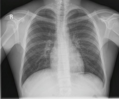
Inclusion
A chest X-ray should include the entire thoracic cage.
Image Quality – Anatomy Inclusion.
- First ribs?
- Costophrenic angles?
- Lateral edges of ribs?
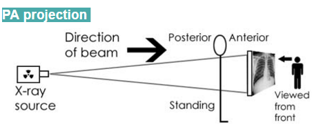
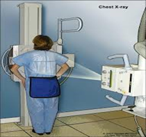
Projection
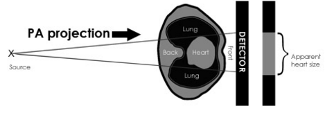
Posterior-Anterior (PA) projection
The standard chest radiograph is acquired with the patient standing up and with the X-ray beam passing through the patient from Posterior to Anterior (PA).
The chest X-ray image produced is viewed as if looking at the patient from the front, face-to-face. The heart is on the right side of the image as you look at it.
Anterior-Posterior (AP) projection
Sometimes it is not possible for radiographers to acquire a PA chest X-ray. This is usually because the patient is too unwell to stand.
The chest X-ray image is still viewed as if looking at the patient face-to-face.

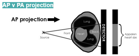
AP v PA – Heart size
The heart, being an anterior structure within the chest, is magnified by an AP view. Magnification is exaggerated further by the shorter distance between the X-ray source and the patient, often required when acquiring an AP image. This leads to a more divergent beam to cover the same anatomical field.
AP v PA – Scapular edges
Radiographers will often label a chest X-ray as either PA or AP. If the image is not labelled, look at the medial edges of each scapula.
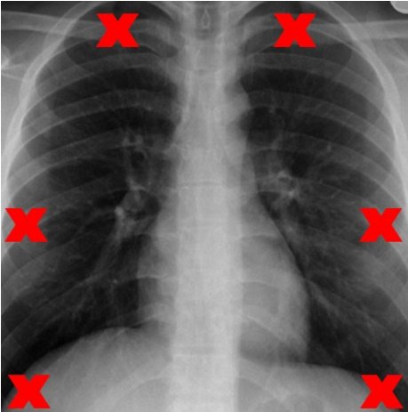
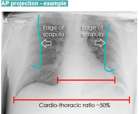
AP projection – Example
- The scapulae are not retracted laterally and they remain projected over each lung.
- Heart size is exaggerated (cardiothoracic ratio approximately 50%).
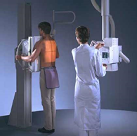
- In order to take a PA view, the patient places his or her arms around the side of the detector plate, or stands with hands-on-hips This ensures the scapulae are rotated laterally and no longer overlap the lungs.

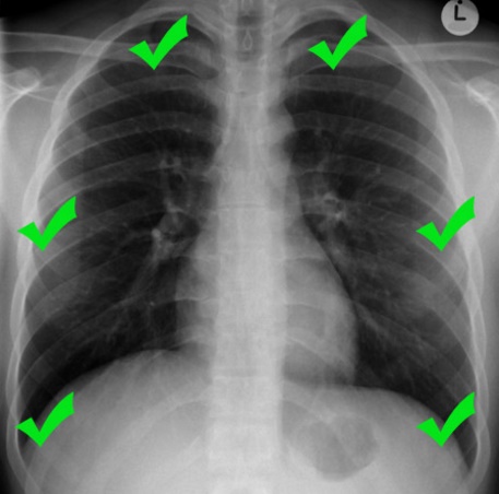
PA projection – Example
The edges of the scapulae are retracted laterally with only a small portion projected over each lung. The lungs are therefore more easily seen.
The cardio-thoracic ratio is clearly well within the normal limit of 50%.
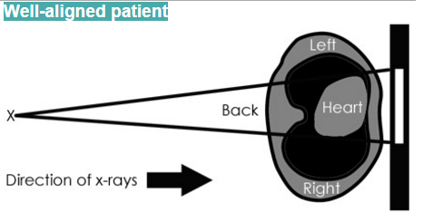
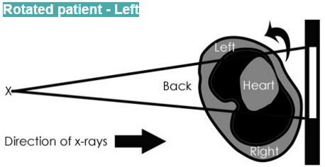
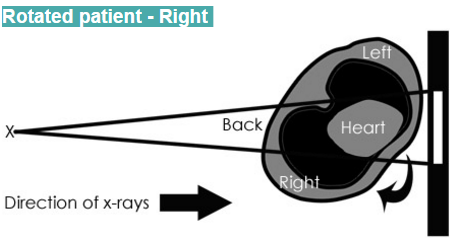
Rotation
If the patient is very rotated and you do not recognize this, certain appearances may become misleading.

Principles of rotation
The spinous processes of the thoracic vertebrae are in the midline at the back of the chest. They should form a vertical line that lies equidistant from the medial ends of the clavicles, which are at the front of the chest.
Does rotation matter?
- It may be difficult to know if the trachea is deviated to one side by a disease process.
- It also becomes difficult to comment accurately on the heart size.
- Changes in lung density due to asymmetry of overlying soft-tissue may be incorrectly interpreted as lung disease.
Inspiration and lung volume
Chest X-rays are acquired in the inspiratory phase of the respiratory cycle. The radiographer asks the patient to, ‘breathe in and hold your breath!’.
Assessing inspiration

To assess the degree of inspiration it is conventional to count ribs down to the diaphragm. The diaphragm should be intersected by the 5th to 7th anterior ribs in the mid-clavicular line. Less is a sign of incomplete inspiration.
Expiration
- Anteriorly only the third rib intersects the diaphragm at the mid-clavicular line.
- The lung bases are white – Is there consolidation?
- How big is the heart?
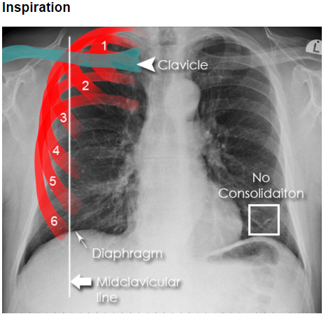
Inspiration
- Anteriorly the sixth rib intersects the diaphragm at the mid-clavicular line.
- The lungs are not consolidated.
- The heart size is clearly normal.
Assessing for Hyper-Expansion
While checking for adequate inspiration you may notice that a patient’s lungs are hyperexpanded (>7th anterior rib intersecting the diaphragm at the mid-clavicular line).
This is a sign of obstructive Airways Disease.
It is possible to assess for Hyper-Expansion by counting ribs, or by checking for flattening of the hemidiaphragms.
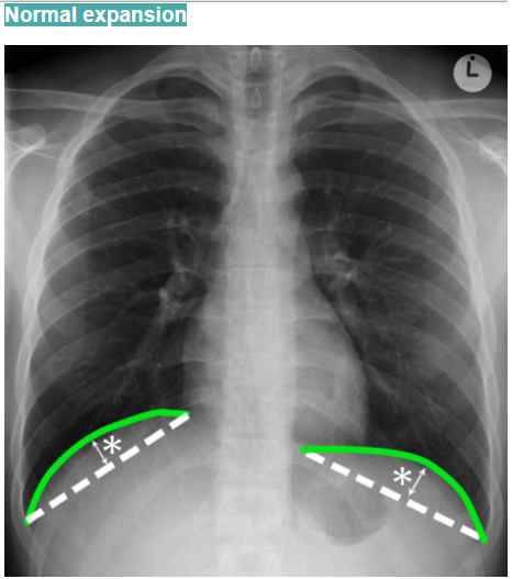
The imaginary dotted line between the costophrenic and cardiophrenic angles.
The distance between this line and the diaphragm (green lines) should be greater than 1.5 cm (asterisk) in normal individuals.
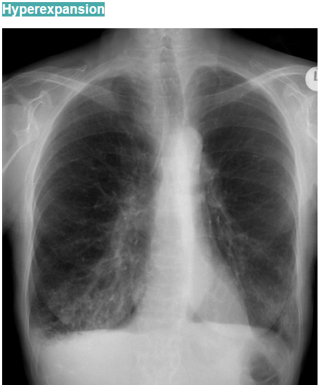
Hyper-Expansion
- These are clearly flattened in this patient.
- The ribs are difficult to count as they have lost density. This is due to long term steroid treatment for the patient’s emphysema. There is also the consolidation of the lung bases due to pneumonia
Penetration
Penetration is the degree to which X-rays have passed through the body.
- The diaphragm is visible to the spine.
- The left paravertebral soft tissues are visible, and the right side of the spine is clear.
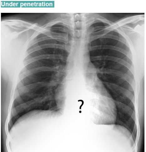
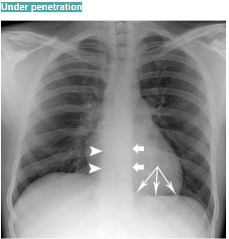
Under penetration
- The left hemidiaphragm is not visible to the spine.
- Lung tissue behind the heart cannot be assessed.
Artifact
Key points
- Some artifacts are unavoidable.
- A chest X-ray may be obtained to assess the position of medical devices.
- Ask yourself if artifact limits image interpretation.
- Can the question clinical question still be answered.
Radiographic Artifact
As previously discussed, examples include rotation, incomplete inspiration and incorrect penetration. Other radiographic artifact includes clothing or jewellery not removed.
Patient artifact
Artifact may be due to patient factors such as poor co-operation with positioning or movement. Very often obesity exaggerates lung density. Occasionally normal anatomical structures such as hair or skin folds can cause confusion.
Download Chest X-Ray Quality: How to Interpret Chest X-Ray PowerPoint File
You May Also Like:
Download Free Chest X-Ray Book: Chest X-Ray Made Easy 4th Edition
